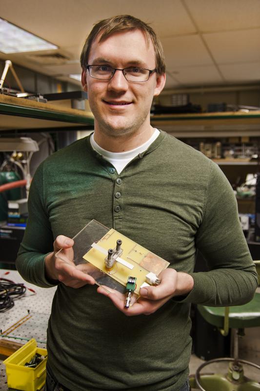
An MU engineering team invented a portable, inexpensive X-ray device with many potential uses in the medical and scientific fields. The team published a paper about the device in the January edition of scientific journal IEEE Transaction on Plasma Science.
The device, which weighs seven grams and is about the size of a stick of gum, is safe for the environment, graduate student and researcher Brady Gall said in an email. It operates on an on-off system, which provides zero risk of radiation contamination, emits low doses of radiation at a safe level for humans and poses no risk to nuclear proliferation. The entire device is operated using a maximum of 10 watts of electrical power.
“The idea for the project sprang from a related, but separate study involving compact ion-thrusters for micro-spacecrafts,” Gall said in an email. “The piezoelectric X-ray source was initially conceived as a diagnostic tool for our particular interests, however, it turned out that it could be generalized for a wider range of applications.”
This minuscule device can turn into an X-ray scanner by attaching it to an X-ray sensitive film or a digital X-ray charge coupled device.
“Our device is analogous to the flash of a camera,” Gall said in an email. “That is to say it provides the light (in our case, X-ray radiation) to illuminate a subject which is then photographed using film or some other means.”
Once the source is converted into a scanner, it can be used in a number of ways, according to an MU News Bureau press release.
Dentists can photograph the insides of patients’ mouths with the X-ray scanner due to its small size. The device uses less energy when attached to rovers used for space exploration, and it can be used to scan cargo for illegal items.
“Current technology requires that a person must stand by the container to probe for nuclear materials, which can prove to be dangerous,” graduate student Emily Baxter said. “This technology is small enough that it could be put in a cargo container and can probe for nuclear materials from within the container.”
Its decreased energy usage can be used in places lacking power, Gall said. Geologists can use it to study minerals in remote areas, like the Sahara Desert, and doctors working in rural areas can use the device to take X-rays of patients.
The X-ray source is built using a safe, man-made ceramic material called lithium niobate, Gall said. The non-radioactive lithium niobate, combined into a crystal, uses a piezoelectric effect to power up the device.
The piezoelectric effect converts electrical energy to mechanical energy and then back to electrical energy, Baxter said.
“We combine both of these effects in a single crystal that converts 12 volts to 120,000 volts,” Gall said. “The trick is that we use a harmonic signal. In layman’s terms, we provide the crystal with a series of electrical ‘kicks.’ This kick makes the crystal expand and contract like a spring. Right when the crystal is about to return to its rest state, we kick it again, this time it stretches out even further.”
The team repeats the effect 3,000 times, creating a 10,000 times increase in voltage, Gall said. An electron accelerator uses this energy to generate X-rays. Gall said the device was successful in producing X-rays and hopes that it will be successful on the market.
The project began in 2007 and was headed by Scott Kovaleski, associate professor of electrical and computer engineering. The team also included graduate students James VanGordon, Emily Baxter, Andy Benwell, Mark Kemp and staff scientist Peter Norgard.
Gall said that the device is still a prototype and will take a few years to be market-ready.
“Well, it is very exciting to be involved in this project and it is also inspiring to see so much interest from the public…it makes me feel proud of our accomplishments,” Gall said. “I am also happy to help promote the University of Missouri as a leader in scientific and engineering innovation.”